


The knee joint arthrosis is a chronic (long -term) degenerative disease that causes cartilage destruction in the joints.Symptoms include pain, stiffness and swelling.Treatment options for reducing pain and disability include lifestyle (diet, physical exercises), physical and professional treatment methods, medicines and surgery changes.
Osteoarthrosis of the osteoporosis of the knee joint
The knee joint osteoarthrosis is a common disease, accompanied by chronic, exhaustive pain.The latest clinical data has shown that central sensitization stimulates the deforming osteoarthrosis of the knee joint.To identify new analgesic goals/new therapeutic strategies, it is essential to understand the arthrosis of the knee joints to influence the central processing of pain.
Inhibitory receptors weaken peripheral immune cells and modulate central neuroimmune responses.The introduction of the receptor agonist systemic weakened the behavior of OA-induced pain and changes in circulating and anti-inflammatory cytokines in this model.
The joint deforming joint
Inflammation and wear on the deforming arthrosis of the knee joint on the bones of the knee joint (osteo = bone, artro = joint, ITIS = inflammation).Diagnosis of osteoarthritis of the knee joint is based on two main results: radiographic data on changes in bone health (using medical images such as X -rays and magnetic resonance) and human symptoms.About 14 million people have symptomatic knee -arthrosis.Although more common in older people, 2 million of the 14 million people were less than 45 of the oa of knees in the diagnosis and more than half were less than 65 years old.
Osteoarthritis (OA knee) is a progressive disease caused by inflammation and degeneration of the knee joint, which deteriorates over time.
This affects the entire joint, including bones, cartilage, ligaments and muscles.Its development is influenced by age, body weight index (BMI), bone structure, genetics, muscle strength and activity.The OA knee can also occur as a secondary condition after the knee injury.Depending on the stage of the disease and the presence of related injuries or conditions, OA knee can be controlled by physical therapy.More severe or expanded cases may require surgical intervention.
Symptoms
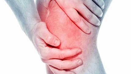
People who develop knee OA may experience a wide range of symptoms and restrictions based on the onset of the disease.Pain occurs when cartilage covering the bones of the knee joint.The areas where the cartilage is worn or damaged explores the underlying bone.The effect of the bone allows you to increase the compression of stress and cartilage and sometimes bone contact during movement, which can cause pain.Because the knee is a joint, activity level, level of activity, and the type and duration of actions generally have a direct effect on the symptoms.Symptoms can deteriorate with weight activity, for example, when they are involved in heavy objects.
You may include the symptoms of the knee joint:
- The deterioration of the pain during or after surgery, especially walking, climbing, walking on the stairs or moving from seat.
- Pain or stiffness after sitting for a long time with a bent or straight knee.Pain is the most common symptom of osteoarthritis.With the development of the disease and inflammation, pain can become constant.
- Moving the knee when you are leaping out, cracks or grinding.
- Swelling after action.
- The stiffness of the affected joint was often seen in the morning and after relaxation.
- Edema, which is sometimes warm, may be noticeable in arthritis.
- Due to osteoarthritis due to bone growth and cartilage loss.The growth of bones in the fingers of the fingers is called Hyberden nodes.Bushar nodes are the growth of bones on the middle joints of the fingers.Degeneration of the cartilage of the knee joint can lead to the outer curvature of the knee (onion foot).
- When the arthritis moves, a cracking sound or grid feeling is noticeable.The reason for this is to delete the bone or coarse cartilage.
Usually, these symptoms do not occur suddenly and at the same time, but gradually develop over time.Occasionally people do not recognize that they have osteoarthritis because they do not remind me of a certain time or injury that caused the symptoms.If the knee pain has deteriorated for several months, which does not react to relaxation or change of activity, it is better to seek advice to a medical worker.
Diagnosis
Osteoarthritis can often be diagnosed with characteristic symptoms of pain, reduced movement and/or deformation.Osteoarthritis can be confirmed by X-ray or MRI scan.General data includes narrowing of joint space between the bones, loss of cartilage and bone drill, or growth of bones.Blood tests can be used to exclude other possible conditions, but they cannot diagnose osteoarthritis.
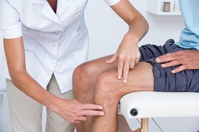
In the knee OA, 2 primary procedures are diagnosed.The first is based on the report on symptoms and clinical trial.The physiotherapist asks questions about medical history and activity.The therapist performs a physical examination to measure knee movement (movement range), strength, mobility and elasticity.They can also seek various movements to see, increase or decrease pain.
The second tool used to diagnose the knee joint is a diagnostic display.The physiotherapist can send you to the doctor, who prescribes the X -Sikra of the knee in a variety of situations to check the damage to the knee joint bone and cartilage.
If you suspect more severe damage to the joints, you can order an MRI to look more closely at the general condition of the joint and surrounding tissues.
Blood tests can also be ordered to exclude other conditions that can cause symptoms similar to osteoarthritis of the knee joints.
Treatment
Depending on the severity of arthritis and the age of the patient, it is chosen to treat the joint joints of the knee joints.Treatment may consist of operational or conservative methods or combinations.
The first treatment line of the knee joint arthritis includes modification of activity, anti -inflammatory drugs and weight loss.
Rejecting pain -enhancing activities can make this condition acceptable to some people.Anti -inflammatory drugs help relieve inflammation, which can contribute to pain.
Physiotherapy to strengthen the muscles around the knee can help absorb the shock of the joint.This is especially true for arthritis with the knee glass (patello-femoral).The load is designed to transfer the load to the part of the knee joint, which is less than arthritis, can also relieve pain.Medicines inside the knee joint can be temporarily helped.
In addition, the opposite side is walking with the reed in his hand, as the painful knee can help distribute part of the load and reduce pain.Finally, weight loss helps to reduce the force passing through the knee joint.A combination of these conservative measures can help relieve pain and prevent disability.
If these methods do not allow the condition to be tolerated, surgery can be the best solution for treating arthritis of the knee joint.The exact type of operation depends on age, anatomy and the main condition.Some examples of surgical options for treating arthritis are osteotomy, which is to adjust the bone cutting of the joint.
Modern methods of treating the arthrosis of the knee joint include osteotomy, which is a good alternative when the patient is young and the arthritis is limited by an area of the knee joint.This allows the surgeon to rebuild the area of the arthritis and not to touch the load relatively on the knee joint.For example, the patient can be rebuilt to redistribute the load through the joint.The advantage of this type of surgery is that the patient's own knee joint is preserved and can potentially ensure the pain for many years to relieve the prosthesis without knee deficiencies.The disadvantages include the longer rehabilitation course and the possibility of arthritis in a recently balanced knee.
The operation of the knee joint replaces the cutting of the arthritic bone and the insertion of the prosthesis joint.All joint surfaces are replaced, including the femur, lower leg and knee cups.The joint surfaces are removed and the end of the bone is replaced by a prosthesis.The component of the prosthesis is usually made of metal and plastic surfaces that are designed to slide smoothly.
The replacement of the knee joint
The general functioning of the knee joint replacement was first performed in 1968, and over the years it has developed a reliable and effective way to get rid of pain and allow patients to continue their active lives.The improvement of surgical methods and implants has contributed to this to be one of the most successful orthopedic procedures.As the population becomes older and remains more active, the need for overall knee replacement continues to increase.Many operations of the knee joint replacement took place in a special surgical hospital.The development of surgical technology and the development of new implants are some contributions to surgeons.
People often think about when and why they should replace their knees.This is an individual issue that depends on the level of human activity and functional needs.Many people with arthrosis are painful that prevent them from participating in activities;Others were so weakened so hard for them to wear shoes and socks.Full replacement of the knee joint offers a solution to the problem of arthrosis and performs pain and activity.After rehabilitation from a successful full exchange of the knee joint, the patient can wait for surgery without pain.Full replacement of the knee joint significantly improves the patient's condition and significantly reduces its long -term treatment costs.This study has shown that not only is the general exchange of the knee joint economically effective, it also provides greater functionality and the best quality of life.
Full replacement of the knee joint is considered a main operation and the solution is not trivial.Usually people decide to go through surgery when they feel they can no longer live with their arthritis.
The implant consists of 4 parts: tibia, femur, plastic insert and pattern.The components of the tibia and the femur are made of metal, usually cobalt chromium, and is used to close the end of the thigh and lower leg after removing the arthritic bone.The plastic insert is made of ultra -high molecular weight polyethylene and fits into the tibia component, so the polished thigh surface slides along the plastic.The component of the knee cup also slides towards the front of the femur.They are usually fixed to the bone cement.
Complete knee replacement in the operating room is carried out with a special laminar airflow system that promotes the likelihood of infection.The surgeon will wear a "space suit" aimed at reducing the likelihood of infection.The whole surgical team consists of the surgeon, two -three assistants and Dada.
The anesthesia is given through an epidural catheter that is a small tube in its back.During surgery, the patient may be awake and sleepy.
After the introduction of the epidural block around the thigh, a tower or cuff is placed.The horizontal rod is overestimated during surgery to reduce blood loss.Cut the knees full of replacement along the front knee.Depending on the anatomy, the incision should be measured between 4 and 10 inches.
The joint surfaces of the femur, lower legs and patellas are exposed and removed using strength tools.At the same time, the knee deformation is corrected and the knee will be straight after surgery.The bone is ready for an artificial knee joint and then places a prosthesis.During closure, two drainage is installed around the workspace to help evacuate blood.SaPers are used to close the skin.
The full operation takes 1-2 hours.The patient was then taken to the recovery room, where the tests are checked.Most patients can be taken to regular rooms for several hours;Others need to stay in the hallway in the cure, as determined by a surgeon and an anesthesiologist.
Patients usually stay in hospital for 3-4 days after full surgery
Risks during surgery
Some risks of the surgical procedure include blood loss, leg formation and probability of infection.The general prevalence of these risks is very small.These should be discussed with the surgeon before starting the surgery.
Some risks of the presence of the prosthesis include the probability that the components may weaken or wear over time or may be infected with the prosthesis.These questions are discussed again with the surgeon.
Post -operation course
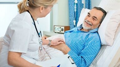
Immediately after a complete operation of the knee joint replacement, the patient falls into the recovery room.Most patients can penetrate a regular ward in a few hours when the sensation returns to the legs.They give the epidural catheter a pain pump that allows you to control the pain.
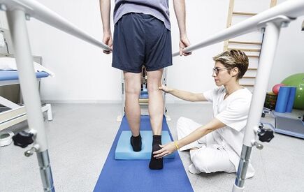
On the day of the operation, you can do some exercises, as indicated by the physiotherapist, including quadruple reduction and moving the legs up and down.Depending on the surgeon's preference, you can start bending the new knee immediately after or on the first day of surgery.The patient is allowed to wet the mouth after surgery, but may drink fluids or cause nausea.The patient will have a catheter in the bladder so you don't have to worry about urination.As soon as the movement is restored, it allows you to sit down, stand up and take a few steps with a pedestrian and therapist.
The first day after surgery will be active and develops to promote more agile.
The patient encounters physiotherapists who teach further exercises.They also help their feet and take a few steps with a walk.As a general rule, the patient allows you to drink clean fluid.
It is easier and easier to move in the next few days.The patient is released from the pain and the urine catheter.The treatment of pain is given in the form of tablets.On the second day after surgery, if signs of healing are found in the intestine, they allow you to eat regular food.
Depending on his age, preoperative physical condition and insurance involvement, the patient may apply for short -term accommodation at a rehabilitation institution.Otherwise, the patient will be allowed at home and the physiotherapist comes to his house to continue rehabilitation.The dispatcher discusses these opportunities with the patient and helps him to plan his return.
The return to the activity is driven by surgeons and therapists.As a general rule, patients can go as much as they want after surgery.Patients can continue to move after 6 weeks.After 8 weeks, patients can continue to play in golf and swimming;They can play tennis every 12 weeks.The surgeon will help you decide what measures can be taken.
What kind of physiotherapist do you need
All physiotherapists make training and clinical experience to treat various conditions or injuries:
- A physiotherapist who has experience in treating the knee joint osteoarthritis and post -surgery to replace the knee joint.Some physiotherapists perform orthopedic focus.
- A physiotherapist who certified an orthopedic clinical specialist.This physiotherapist will have advanced knowledge, experience and skills that can be applied in a state.
- You can find physiotherapists who have these and other accounting data with the help of an MRI, an online device that promotes the determination of physiotherapists with specific clinical knowledge.
General advice if you can find a physiotherapist (or any other medical service provider supplier):
- Recommendations from family and friends or other suppliers of medical services;
- Turning to the admission physiotherapy clinic, you must ask physiotherapist experience to help people with arthritis.
During the first visit of the physiotherapist, you should prepare for a more detailed description of the symptoms and report on the activities that exacerbate the condition.